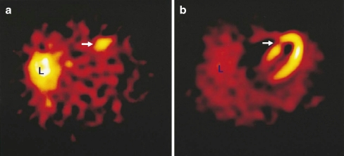
Fig. 2.
Transverse tomographic images in a patient with acute anteroseptal infarction. a Arrow shows increased 99mTc-labelled Anx A5 uptake in the anteroseptal region 22 h after reperfusion. b Perfusion scintigraphy with sestamibi 6–8 weeks after discharge shows an irreversible perfusion defect which coincides with the area of increased 99mTc-labelled Anx A5 uptake (arrow). L liver. Reprinted with permission from Elsevier (The Lancet, 2000, 356, 211)

