Fig. 5.
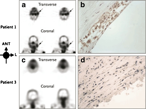
Images of unstable atherosclerotic carotid artery lesions obtained with radiolabelled Anx A5. a Transverse and coronal views obtained by SPECT in patient 1, who had a left-sided TIA 3 days before imaging. Although this patient had clinically significant stenosis of both carotid arteries, uptake of radiolabelled Anx A5 is evident only in the culprit lesion (arrows). b Histopathological analysis of an endarterectomy specimen from patient 1 (polyclonal rabbit anti-Anx A5 antibody, ×400) shows substantial infiltration of macrophages into the neointima, with extensive binding of Anx A5 (brown). c In contrast, SPECT images of patient 3, who had had a right-sided TIA 3 months before imaging, do not show evidence of Anx A5 uptake in the carotid artery region on either side. Doppler ultrasonography revealed a clinically significant obstructive lesion on the affected side. d Histopathological analysis of an endarterectomy specimen from patient 3 (polyclonal rabbit anti-Anx A5 antibody, ×400) shows a lesion rich in smooth muscle cells, with negligible binding of Anx A5. ANT anterior, L left. Copyright © 2004 Massachusetts Medical Society. All rights reserved. [43]

