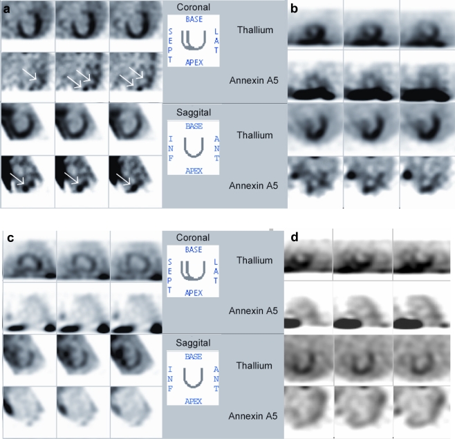
Fig. 6.
Dual-isotope imaging using 201Tl for left ventricular contour detection and, simultaneously, radiolabeled annexin A5 in patients with dilated cardiomyopathy. a Dilated cardiomyopathy patient with rapid deterioration of left ventricular function. Note focal uptake in apex and lateral wall, and slight septal uptake. b Dilated cardiomyopathy patient in acute heart failure. Note global uptake of radiolabeled annexin A5. c Dilated cardiomyopathy patient in stable clinical condition. Uptake is absent even when image is enhanced to the extent that background radioactivity can be observed. d Family member of patient in panel b. No clinical evidence is seen of dilated cardiomyopathy. Note absence of uptake of radiolabeled annexin A5. ANT, anterior; INF, inferior; LAT, lateral; SEPT, septal. Reprinted by permission of the Society of Nuclear Medicine from [90]

