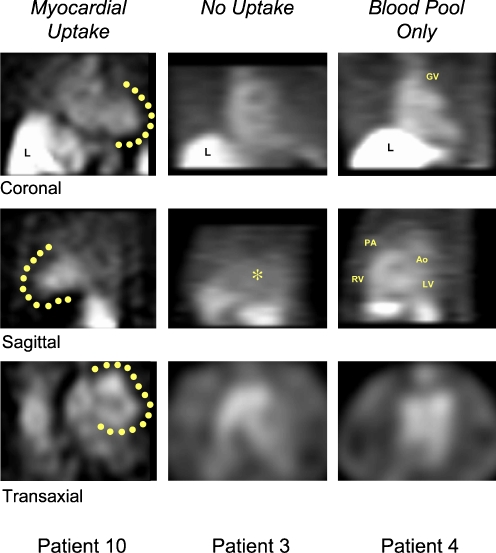
Fig. 7.
SPECT images of 99mTc-labelled Anx A5 in three patients (10, 3 and 4) are demonstrated. a In patient 10, myocardial uptake (as outlined by solid circles) is clearly seen in all tomographic orientations and can be differentiated from the left ventricular cavity, especially in the transaxial slice. b In patient 3, by contrast, no Anx A5 uptake is observed, and only background activity is seen. c Patient 4 demonstrates Anx A5 activity in the blood pool originating from the great vessels (GV great cardiac vein, PA pulmonary artery, Ao aorta) and ventricular contours, but no activity is observed in the myocardium. The scan of patient 10, with myocardial uptake, was further processed (Fig. 8). RV right ventricle, LV left ventricle. Adapted by permission from Macmillan Publishers Ltd: Nature Medicine [42], copyright 2001

