
Fig. 3.
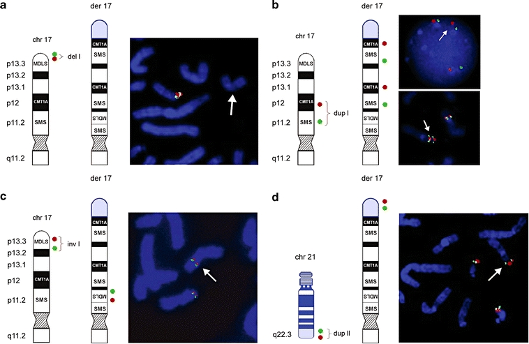
Ideograms and FISH results of patient 1. Schematic representation of a normal 17p and der(17) (black and white) with translocated chromosome 21 material (blue). The location of the FISH probes are shown on the left side of each figure panel; der(17) is indicated on FISH pictures by a white arrow. a Terminal deletion of 17pter was validated using BAC clones RP11-1260E13 (red) and CTD-2326F1 (green) (del I). b FISH with PMP22-specific PAC RP1-150M12 (red) RA11-specific and BAC RP11-525O11 (green) revealed direct duplication of the CMT1A and SMS regions in 17p12p11.2 (dup I). c FISH with PAC RP1-95H6 (red; adapted from Chong et al. 1997) and BAC GS-202L17 (green; adapted from Knight et al. 2000) showed inverted insertion of the MDLS region into the SMS region. d Array CGH also identified a duplication of 21q22.3 (dup II). Additional FISH analysis using BAC clones RP11-40L10 (green) and RP11-16B19 (red) revealed that the duplicated material of 21q22.3 was translocated onto der(17). Summary of FISH results is provided in Table 1