
Fig. 4.
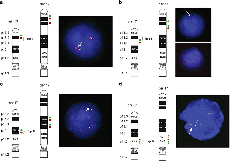
Ideograms and FISH results of patient 2. Schematic representation of the normal 17p and der(17). The locations of the FISH probes are shown on the left side of each figure panel. a The distal breakpoint of duplication I showed a relatively simple fluorescence signal pattern with probes RP11-810M2 (green; normal) and RP11-597I9 (red; duplicated). b The proximal breakpoint of duplication I showed a duplicated signal for RP11-222J21 (green) and a normal signal for RP11-98D15 (red). c Direct orientation of duplication III was shown using BAC clones RP11-601N13 (green) and RP11-726O12 (red). d For the distal breakpoint of duplication IV, BAC clones RP11-448D22 (green) and CTD-2145A24 (red) showed duplicated signals on der(17), indicating that both middle SMS-REP and LCR17pB are duplicated as a block. Four red signals on der(17) representing two normal and two duplicated copies of LCR17pA/B (dup III) and LCR17pB (dup IV) and four green signals depicting three normal copies of SMS-REPs and the duplicated middle SMS-REP. Summary of FISH results is provided in Table 2