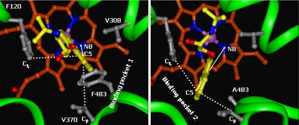
Fig. 3.

Representative snapshots of two distinct binding modes observed in simulations of WT CYP2D6 (binding mode 1, left panel) and the F483A mutant (binding mode 2, right panel). Propranolol is depicted in yellow sticks and hydrophobic substrate binding amino acid residues are depicted in grey ball-and- sticks. Two geometric features used to discriminate between the two binding modes are displayed: the distance between the nitrogen atom of heme pyrrole ring B (NB) and the C5 atom of R-propranolol and the angle defined by the Cξ atom in F120, the C5 atom in R-propranolol and the Cβ in V370
