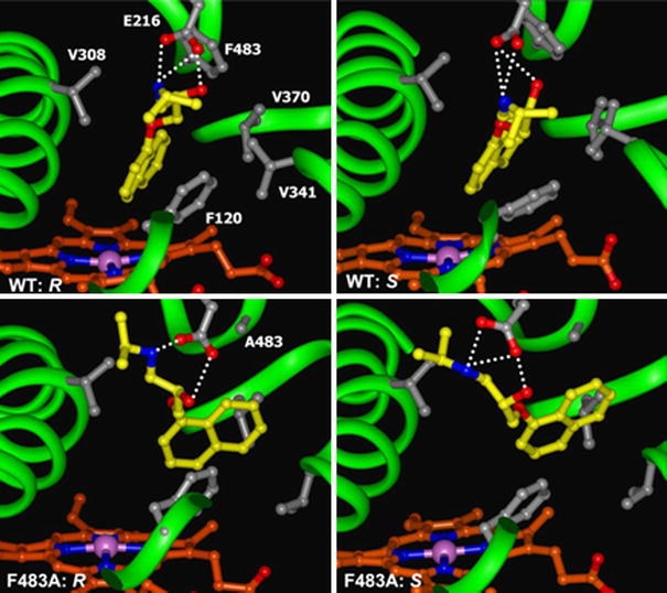
Fig. 5.

Representative orientations of R- and S-propranolol in the binding cavities of WT and mutant CYP2D6 observed during the MD-simulations at points defined in Fig. 1 and included in approach 2: e (WT: R), g (WT: S), i (F483A: R), and k (F483A: S). R-propranolol forms fewer hydrogen bonds than S-propranolol in the F483A mutant. The F483A mutation also causes a loss of favourable hydrophobic interactions, which can be compensated by increased hydrogen bond formation by S-propranolol, but not by R-propranolol
