
Figure 7.
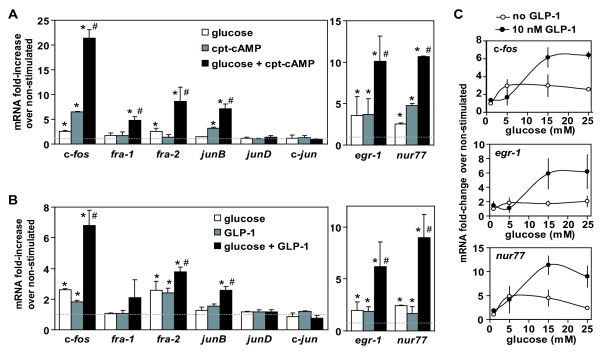
Induction of IEGs by metabolic stimuli in isolated rat islets. Rat islets were isolated, cultured and serum deprived at reduced glucose concentration (1 mM) for 20 hours. Stimulation was done for one hour with 0.2 mM cpt-cAMP and/or 25 mM glucose (A); or with 10 nM GLP-1 and/or 25 mM glucose (B). mRNA levels for mentioned genes were quantified in triplicate by real-time RT-PCR, normalized with reference to 18S rRNA, and are shown as fold-increase over non-stimulated controls. Shown are the means of values obtained for three (A) or two (B) independent experiments (error bars = s.d.). Student T-tests were used for statistical analysis; *p < 0.05 vs non-stimulated; # p < 0.05 vs single stimulus conditions. C) Effect of various glucose concentrations on the induction level of IEG expression (after one hour stimulation). Results as mean of at least two independent experiments (s.d. as error bars).