
Figure 7.
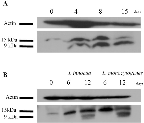
Kinetic of granulysin processing in LAK and Listeria specific T cells. LAK (A) or antigen specific T cells (B) were taken out of culture at indicated time points followed by lysis of cells and separation of proteins by SDS-PAGE. Proteins were blotted and detected using anti-granulysin Ab (1:1000). The blot was stripped and probed for the β-actin.