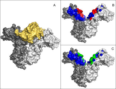
Figure 1. Protein–Protein Interfaces, Hotspots, and Predictions.
Residues that are part of protein–protein interfaces often constitute a large fraction of the protein. Hotspot residues, namely residues that upon mutation hamper the interaction, are only a small fraction of these interface residues. Interestingly, methods designed to predict interface residues usually capture only a small fraction of them.
(A) Human growth hormone (yellow) bound to the extracellular portion of its homodimeric receptor.
(B) The chains of the receptor (gray) are 201 residues long. The protein–protein interface covers 31 of these residues (blue and red) on each of the chains. Mutating one of the six residues colored in red abrogates or severely hampers the interaction.
(C) A prediction method (ISIS; see text) that was designed to identify all interface residues managed to capture only five of the interface residues (colored green).

