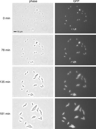
Figure 2. Viability of printed E. coli.
A timed progression of phase contrast microscopy images shows cells printed in a single droplet growing over 3 hours at 30°C. The corresponding fluorescence images show that all the cells in this droplet are viable. The overall viability was greater than 98.5% (see text).

