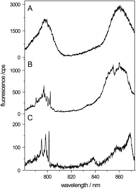
FIGURE 2.
Low-temperature fluorescence-excitation spectra of membrane-reconstituted LH2 from Rps. acidophila. The spectra were taken of samples with a protein/lipid-ratio (w/w) of (A) 1:50, (B) 1:40000, and (C) 1:160000. Decreasing the protein/lipid ratio corresponds to less LH2 complexes per vesicle and for a ratio of 1:160000 on average less than one complex per vesicle can be expected. This is reflected in the spectrum (C), which exhibits typical features of a single-complex spectrum. The spectra were measured in the confocal mode of the setup. Further details are given in the text.

