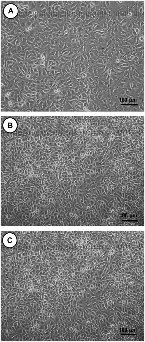
FIGURE 10.
Light microscope images of (A) B16F10 cells grown in normal CM on a culture dish; (B) the B16F10 cells with PEG43 after growing a further 24 h. Here, the B16F10 cells were allowed to grow for 24 h in a CM with PEG43, and washed with PBS. Normal CM without PEG43 was then added and the photo taken. (C) The B16F10 cells without PEG43 after growing a further 24 h. Here, the B16F10 cells were allowed to grow for 24 h in normal CM, and washed with PBS. Normal CM without PEG43 was then added and the photo taken.

