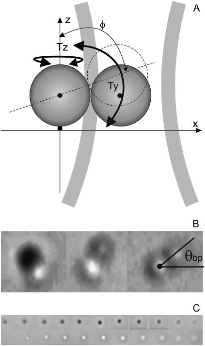
FIGURE 2.
(A) Schematic diagram of a bead pair attached to a surface by a motor at x = y = z = 0 and held by the optical trap. (B) Bright-field video microscope images of bead pairs with different values of the angle φ as defined in panel A. We chose pairs with φ ∼ 90° like the one in the right panel, for which the angle θbp is defined. (C) Bright-field images of a single bead scanned in 100-nm steps in z. The accuracy with which z = 0 could be set by eye using video images was found to be ∼30 nm.

