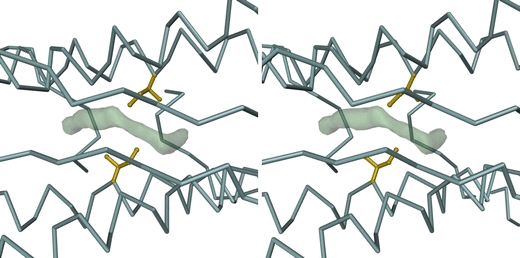
Fig. 3.

Stereoview of a dimer interface in the as-isolated PfFtn structure, showing a difference electron density feature, drawn at the 2.5 map root mean square (RMS) level, which results from a disordered chain of water molecules trapped within a hydrophobic pocket centered around Ile51. This feature is observed in all dimers of all PfFtn structures investigated. The bulk of the PfFtn dimer is represented by its Cα trace (blue-gray). The side chains (including Cα atoms) of the Ile51 residues in both monomers are represented in ball-and-stick mode and are colored gold. The view is down the noncrystallographic twofold symmetry axis of the dimer, looking towards the inside of the PfFtn 24-mer. The figure was prepared with DINO [52]
