Fig. 4.

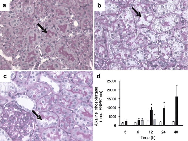
Proximal tubular damage during endotoxemia. Histological examination after LPS+ revealed damage to the proximal tubules compared to LPS−. a LPS−-treated rat with intact brush border membrane in proximal tubules. b Twelve hours after LPS+, damage to the proximal tubules is observed with loss of brush border membranes and formation of vacuoles. c Treatment with both LPS+ and aminoguanidine resulted in less damage to the proximal tubule. Kidneys of LPS+-treated rats killed after 24 or 48 h also showed signs of proximal tubule damage (data not shown). Proximal tubules are indicated by arrows. Original magnifications (a–c) ×400. d Histological data are supported by an increase in the activity of alkaline phosphatase, marker for proximal tubule damage, in urine samples 12 h after LPS+ administration (closed bars, n = 6), compared to controls (open bars, n = 3). Coadministration with aminoguanidine (gray bars, n = 6) reduced this proximal tubule damage. Data are expressed as mean±SE. Significantly different compared to the LPS− (double asterisksP < 0.01) or LPS+ (sharp signP < 0.05)
