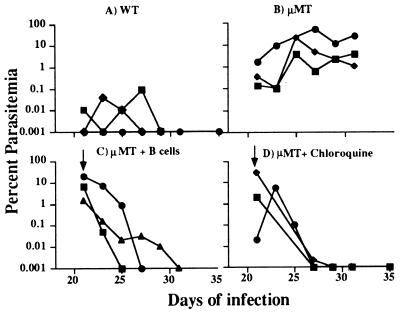
Figure 1.
Course of a primary P. chabaudi infection in female WT (A) and μMT mice (B) and in μMT receiving immune B cells (C) or chloroquine and pyrimethamine (D). Each line represents the infection in an individual mouse. The arrow indicates the time of adminstration of immune B cells (2 × 10⁷ given intravenously) or chloroquine and pyrimethamine (as described in text).

