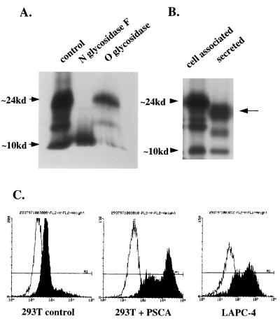
Figure 4.
Biochemical analysis of PSCA. (A) PSCA was immunoprecipitated from 293T cells transiently transfected with a PSCA construct and then digested with either N-glycosidase F or O-glycosidase. (B) PSCA was immunoprecipitated from 293T transfected cells and from conditioned medium from these cells. Cell-associated PSCA migrates higher than secreted PSCA on a 15% polyacrylamide gel. (C) Flow cytometry analysis of mock-transfected 293T cells, PSCA-transfected 293T cells, and LAPC-4 prostate cancer xenograft cells by using an affinity-purified polyclonal anti-PSCA antibody. Cells were not permeabilized to detect only surface expression. The y axis represents relative cell number and the x axis represents fluorescent staining intensity on a logarithmic scale.

