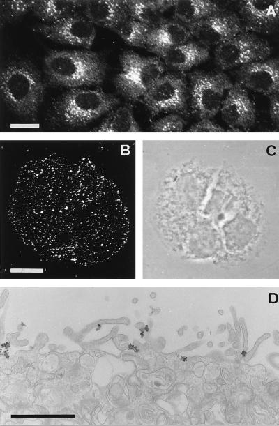
Figure 1.
Immunocytochemical localization of endogenous APP in FRTL-5 cells (A) and of recombinant sAPP (B and D). Visualization of endogenous APP with an antibody directed against the cytoplasmic C-terminal domain (antiserum 2189) resulted in a characteristic crescent perinuclear staining indicative of a Golgi localization of APP (A). For visualization of recombinant sAPP, cells were incubated with 100 nM recombinant His-tagged sAPP for 20 min at 4°C, i.e., under conditions that halted membrane flow. The recombinant sAPP was immunolabeled with specific antibodies recognizing only recombinant sAPP, but not endogenous APP (antibody 3329; see Fig. 2), and detected with either fluorescently labeled (B) or gold-conjugated antibodies followed by silver enhancement (D). The phase-contrast image corresponding to B is shown in C. [Bars = 10 μm (A and B) and 2 μm (D).]

