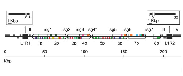
Figure 1.

Structural organization of GiBV proviral locus 1. Proviral genome segments are labeled 1p-8p, with the square and pointed ends representing the 5' and 3' ends, respectively, relative to the putative excision motif. Inter-segmental regions are labeled isg1-isg7, and sequence regions outside the proviral genome segment sequences are labeled I-IV. The flanking tandem repeat regions (solid black squares) are labeled L1R1 and L1R2, and their structure is shown in the open boxes as black boxes in parentheses followed by the copy number of repeat as a subscript. The 2 BAC sequences were joined in isg4 (*) allowing the entirety of each proviral segment sequence to originate from a single BAC clone. Colored boxes represent genes; grey boxes are non-packaged genes, light green boxes are hypothetical proteins without gene family assignment, and the remaining colors represent different gene families.
