Figure 1.

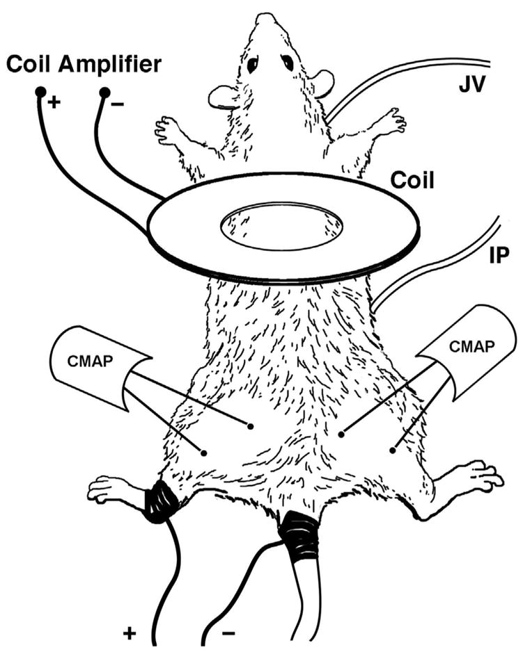
Illustration depicting anesthetic and treatment injection sites (IP, JV respectively), hind limb electroporation path (dark areas of tail and left leg), and CMAP stimulation (Coil) and recording sites (CMAP) on anesthetized rat.

Illustration depicting anesthetic and treatment injection sites (IP, JV respectively), hind limb electroporation path (dark areas of tail and left leg), and CMAP stimulation (Coil) and recording sites (CMAP) on anesthetized rat.