
Fig. 2.
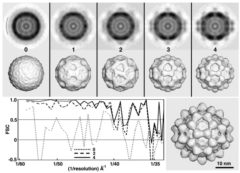
Test of RM method with SBNV images. Central sections (top row; darker shades correspond to higher particle densities) and shaded-surface representations (2nd row) computed from 3D maps corresponding to MODELrand (cycle 0) and MODELsrch(1-4) (cycles 1–4). All 3D maps shown in the top two rows were corrected for effects of the microscope CTF as described in Methods and were computed to a resolution limit of 35 Å. The black and white arcs in the first central section (top left) identify the upper and lower limits of particle radii included in the calculations. FSC plots (lower left) provide a quantitative measure of the quality of each search map. Lower right: CTF-corrected, 3D cryo-EM reconstruction of SBNV, computed to 25Å resolution from 1500 particle images extracted from 29 micrographs.