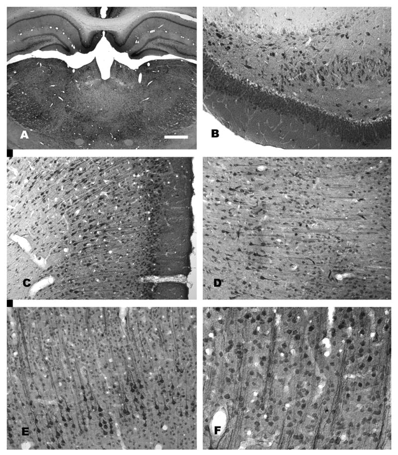
Figure 3.

NAA-immunoreactivity in the rat forebrain. A low magnification photomicrograph of NAA staining in the thalamus and hippocampus is shown in (A). Staining is stronger in most gray matter areas as compared with white matter. Immunoreactivity in the hippocampus is strongest in pyramidal cells, polymorph cells and granule cells (B). Strong NAA-IR is also observed in cortical areas including retrosplenial granular cortex, where both pyramidal and granule cells are strongly immunoreactive (C). In neocortex, staining is particularly strong in layer 5 pyramidal cells, such as those in temporal cortex (D) and motor cortex (E). The columnar organization in these cortical areas can be discerned in NAA-stained sections wherein vertical columns of clustered apical dendrites stained for NAA can be seen (D–F). For methods, see (Moffett and Namboodiri, 1995). Bar = 600 μm A, 100 μm B-E, 50 μm F.
