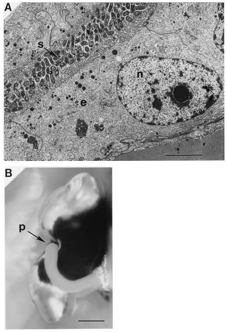
Figure 1.
The light organ environment of symbiotic V. fischeri cells. (A) Thin-section transmission electron micrograph showing symbionts (s) colonizing a portion of the light organ crypts of an adult E. scolopes. The crypts are bounded by microvillus epithelial cells (e), which are believed to supply nutrients to the symbiont population (12). A host cell nucleus (n) can also be seen. (Bar = 5 μm.) In an adult animal, all of these crypts join at and exit through two lateral pores, one on each of the lobes (12). (B) One lobe of the light organ of an adult E. scolopes squid viewed during the process of venting. The crypt contents can be seen exiting the pore (p) of the light organ, appearing as a whitish cylindrical stream. (Bar = 1 mm.)

