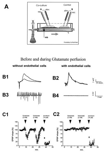
Figure 2.
(A) Scheme of the experimental electrophysiological set-up. A coculture (A) or a control culture (B) were simultaneously stimulated and recorded in an interface-type chamber. Perfusion medium flows beneath the membrane insert (mb). (B1 and B2) Representative field potentials recorded in the pyramidal layer before and after Glu perfusion in control cultures and cocultures. Note the important decreasing in EPSP responses occurring for Glu perfusion in control cultures (B1). Rhythmic activity was observed when recording spontaneous activity during Glu perfusion (B3). No modification of either evoked responses (B2) or spontaneous activity (B4) was observed in cocultures (n = 4). (C1) Synaptic responses (slope of EPSP, percent) in control cultures (n = 4) before and during perfusions of Glu and picrotoxin. EPSP decreasing was observed only when picrotoxin was perfused in coculture experiments (C2).

