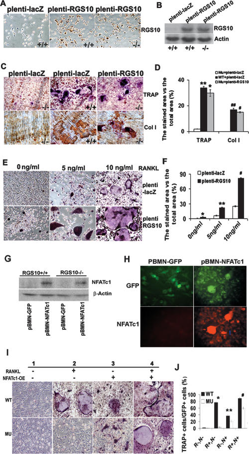
Figure 6.
Rescue of RGS10−/− osteoclast differentiation by reintroduction of RGS10 and NFATc1 and increase in sensitivity of osteoclast differentiation to RANKL signaling by RGS10 overexpression. (A) The BMMs from RGS10+/+ and RGS10−/− mice were infected with plenti-LacZ and plenti-RGS10. Immunostaining results showed that 90% of the cells expressed RGS10 in plenti-RGS10-infected RGS10+/+ and RGS10−/− cells. (B) Western blot results confirmed the expression of RGS10 in plenti-RGS10-infected RGS10+/+ and RGS10−/− BMMs. (C) The BMMs from RGS10+/+ and RGS10−/− mice were infected with plenti-LacZ as a positive control and negative control, respectively. plenti-RGS10-infected BMMs and two controls were induced with RANKL/M-CSF as described in Materials and Methods. The cells were stained for TRAP activity. Note that some TRAP+ cells are multinucleated in plenti-RGS10-transfected RGS10−/− cells and have bone resorption activity on dentine slices (immunostaining of anti-collagen I protein; bone resorption areas become brown or dark brown), indicating that overexpression of RGS10 could rescue osteoclastogenesis. (D) Quantitative analysis of TRAP or collagen I-positive area in C expressed as the percentage of the positive stained area versus total area. Data are presented as mean ± SD. N = 3. For TRAP staining, P < 0.001 (*), Mu-plenti-LacZ versus Mu-plenti-RGS10; P > 0.05 (**), WT-plenti-LacZ versus Mu-plenti-RGS10. For immunostaining of collagen I protein, P < 0.001 (#), Mu-plenti-LacZ versus Mu-plenti-RGS10; P > 0.05 (##), WT-plenti-LacZ versus Mu-plenti-RGS10 (ANOVA). (E) The BMMs from wild-type mice were infected with plenti-LacZ or plenti-RGS10 and then treated with 0, 5, and 10 ng/mL RANKL in the presence of 10 ng/mL M-CSF for 96 h. (Bottom left panel) Without RANKL induction, 8% of precursor cells differentiated into mononuclear TRAP+ cells in plenti-RGS10-infected cells. In the presence of 5 or 10 ng/mL RANKL and 10 ng/mL M-CSF (middle and right panels), there are 3.6-fold and 3.2-fold more mononuclear and mature multinuclear TRAP+ cells, respectively, in the plenti-RGS10 group (bottom panels) compared with the plenti-LacZ group (top panels). (F) Quantitative analysis of TRAP+ cells in E. N = 3 (student’s t-test). plenti-LacZ versus plenti-RGS10 at 0 ng/mL ([*] P < 0.05), 5 ng/mL ([**] P < 0.001), and 10 ng/mL ([#] P < 0.001). (G) Western blot of NFATc1 protein in RGS10+/+ and RGS10−/− BMMs expressing pBMN-NFATc1 or control pBMN-GFP. Overexpression of NFATc1 rescues its expression in RGS10−/− BMMs. (H) NFATc1 and GFP expression in BMMs transfected with pBMN-NFATc1 or pBMN-GFP. Ninety-eight percent of transfected cells become GFP+ cells. pBMN-NFATc1 transfection induces expression of NFATc1 without RANKL induction. (I) TRAP stain of wild-type and RGS10 mutant (MU) BMMs with (panels 2,4) or without (panels 1,3) RANKL induction and with (panels 3,4) or without (panels 1,2) transfection with pBMN-NFATc1. (Bottom, panels 3,4) Overexpression of NFATc1 rescues osteoclast formation with or without RANKL induction. (J) Quantitative analysis of TRAP+ cells in I. Data are presented as mean ± SD. N = 3. (*) P < 0.001, WT−R+,N− versus Mu− R+,N−; (**) P < 0.05, WT−R−,N+ versus Mu− R−,N+; (#) P < 0.05, WT−R+,N+ versus Mu− R+,N+. (R) RANKL; (N) overexpression of NFATc1; (+) presence; (−) absence.

