
Fig. 1.
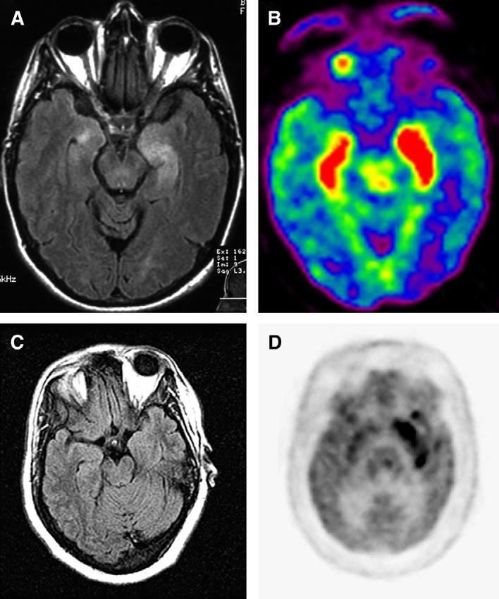
MRI and FDG-PET in patients who have paraneoplastic limbic encephalitis (A) (brain MRI) and (B) (FDG-PET) of a patient who had SCLC and anti-Hu associated paraneoplastic limbic encephalitis; note that there is bilateral medial temporal lobe FLAIR hyperintensity that correlates with FDG hyperactivity (red) in the PET study. (C) Brain MRI and (D) FDG-PET of a patient who had paraneoplastic encephalitis associated with carcinoma of the thymus; in this patient the PET study revealed an extensive area of FDG hyperactivity (dark signal in the medial left temporal lobe) that was not noted in the MRI.