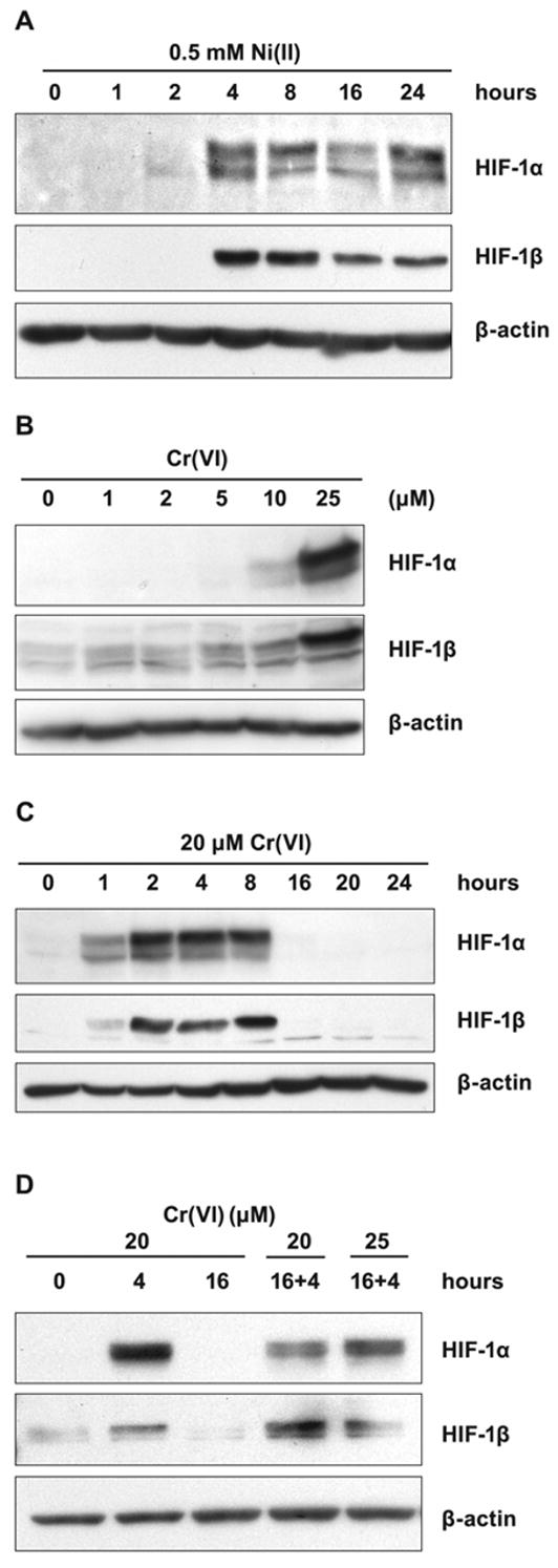
Figure 4.

Effects of Ni(II) exposure on HIF-1α and HIF-1β levels in A549 cells. A, A549 cells were exposed to 0.5 mM NiSO4 for the time periods shown in the Figure. Nuclear protein extracts (15 μg) were prepared for immunoblotting as described in Materials and Methods and probed with antibodies directed against HIF-1α and HIF-1β. The membrane was also probed with antibodies against β-actin to provide loading control. B, A549 cells were exposed for 4 h to various concentrations of K2CrO4 shown in the Figure. The assay was carried out as described in A. C, A549 cells were exposed to 20 μM K2CrO4 for the time periods shown in the Figure. The assay was carried out as described in A. D, A549 cells were exposed to 20 μM K2CrO4 for 4 or 16 h, after 16 h fresh aliquots of K2CrO4 were added to make 20 and 25 μM for an additional 4 h. The assay was carried out as described in A.
