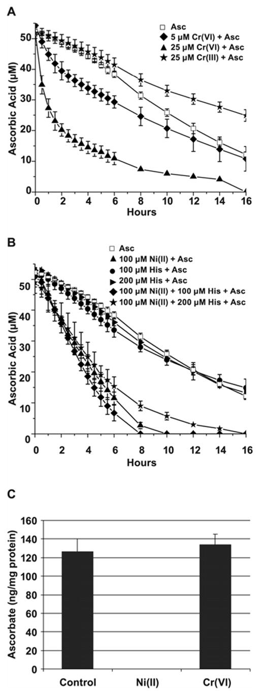
Figure 5.

Kinetics of ascorbate oxidation in the presence of metal ions. A, Ascorbate oxidation by Cr(VI) or Cr(III) in cell-free system. 50 μM of ascorbate in Hepes buffer was incubated at 37ºC alone or in the presence of 25 μM Cr(III), or 5 μM, or 25 μM Cr(VI) for the time periods shown in the Figure. The data are presented as mean values ± the SD. B, Ascorbate oxidation by Ni(II) in cell-free system. 50 μM of ascorbate in Hepes buffer was incubated at 37ºC alone or in the presence of 100 μM Ni(II), 100 μM or 200 μM histidine, or all together for the time periods shown in the Figure. The data presened are mean values ± the SD. C, Ascorbate oxidation by metals in 1HAEo- cells. The cells were preloaded with 50 μM ascorbate for 2 h followed by incubation with 0.5 mM NiSO4 or 5 μM K2CrO4 for additional 16 h. Levels of reduced ascorbate were measured at the end of incubation using HPLC as described in Materials and Methods. The data are presented as means ± SD.
