
Figure 7.
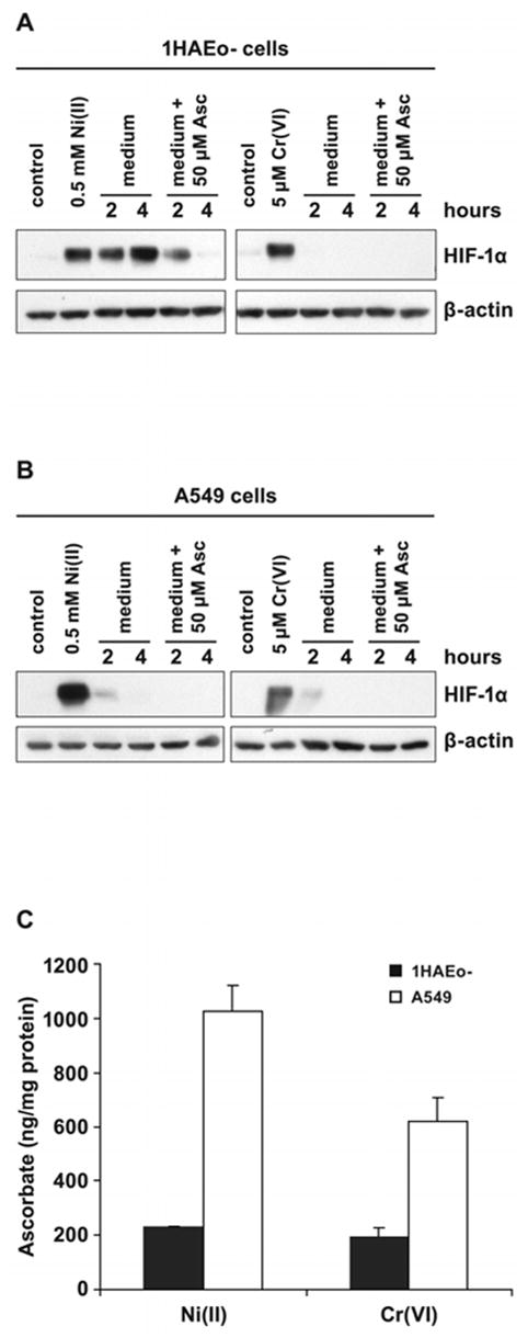
The addition of ascorbate destabilizes metal-induced HIF-1α protein. A, 1HAEo- cells were exposed to 0.5 mM NiSO4 or 5 μM K2CrO4 for 6 h.. B, A549 were exposed to 0.5 mM NiSO4 and 20 μM K2CrO4 for 6 h. The medium was changed to the fresh medium with or without 50 μM ascorbate for the time periods shown in the Figure. Nuclear protein extracts (15 μg) were prepared for immunoblotting as described in Materials and Methods and probed with antibodies against HIF-1α and also probed with antibodies against β-actin to provide loading control. C, The intracellular accumulation of reduced ascorbate following Ni(II) or Cr(VI) exposure. 1HAEo- cells (dotted bars) were exposed to 0.5 mM NiSO4 or 5 μM K2CrO4 for 6 h, or A549 cells (striped bars) were exposed to 0.5 mM NiSO4 or 20 μM K2CrO4 for 6 h, then washed and loaded with 50 μM ascorbate. The intracellular ascorbate level was determined at 2 h of incubation with ascorbate using HPLC as described in Materials and Methods. The data are presented as means ± SD.