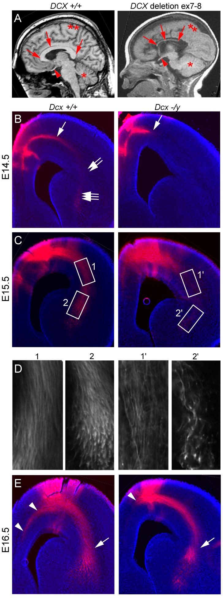
Figure 1.

Delayed axonal extension in Dcx −/y brains. (A) Midline sagittal T1-weighted brain MRI from normal showed well-formed CC and a male with deletion of DCX exon 7-8 showed severe CC hypoplasia (arrows). Optic nerve (arrowhead), cerebellum (*), cortex (**). (B) E14.5 DiI injected into medial subcortical region showed extensive fibers in subcortical white matter (arrow), cortico-striatal (CS) boundary (double arrows) and striatal-thalamus region (triple arrows). Mutant showed minimal axonal extension from the injection. (C) E15.5 DiI injection showed labeling of corticothalamic (CT) axons at CS boundary (box 1) and striatal-thalamus region (box 2), whereas mutant showed diminished labeling. (D) High power views. (E) E16.5 DiI injection showed diminished axon extension of the CC tract (arrowhead) in mutant. There was some catch-up extension of CT tract by this age (arrow).
