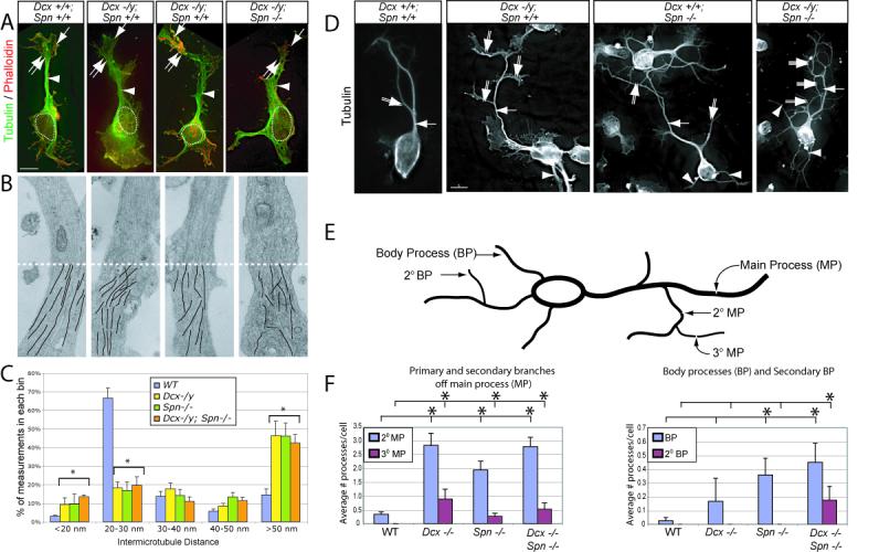
Figure 3.

Disrupted shaft MT cytoskeleton in Dcx, Spn and DKO mutant neurons results in excessively branched neurite phenotype. (A) WT showed condensed MTs in the shaft (arrowhead). Wrist (double arrow) and growth cone (arrow) are well-delineated from the cell body (dashed circle = nucleus). In Dcx −/y or Spn −/− neurons, MTs were instead splayed (arrowhead), and as a result, the neurite shaft width was increased. (B) Transmission electron microscopy showed parallel MT arrays that maintained relatively consistent spacing along the shafts in cultured cortical neurons in wt littermates. This array was disrupted in both single knockouts and was more severe in the DKO. Bottom half (dashes) of each shows line tracings depicting MTs. 8000X. (C) Binned inter-MT distance between nearest neighbor was significantly greater in mutants. N = 1499 total measurements, from 9 wt, 5 Dcx−/−, 4 Spn −/− and 5 DKO neuronal shafts from two separate culture experiments. Error bar = SEM. * = p < 0.05, Chi squared contingency table. (D) WT neurons typically displayed a single monopolar main process (arrow) with occasional 2° branches (double arrow) after 36 hrs in culture. Both Dcx −/y and Spn −/y neurons exhibited excessive 2° (double arrows) and 3° (triple arrows) branches from the primary neurite, as well as an increased number of processes extending from the cell soma (arrowhead). This was most striking in DKO. (E) Quantification of neurite branching. Branches from the main process (MP) were termed 2° MP, and branches from 2° MP were termed 3° MP. Neurites extending from the soma were termed body processes (BP), and branches from the BP were termed 2° BP. (F) WT cells typically had a single MP, with average number of 2° MP per cell less than 0.5. The branching and frequency of 3° MP was increased in Dcx −/y, Spn −/− and DKO neurons. Furthermore, 2° BP were only noted in DKO neurons. * = p < 0.05, pairwise comparison, Student t-test.
