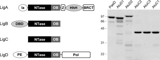
Figure 1.
Multiple DNA ligases of A. tumefaciens. The Atu LigA, LigB, LigC and LigD polypeptides are depicted (left panel) in cartoon form with the N-termini on the left and the C-termini on the right. The core ligase catalytic domains, composed of nucleotidyltransferase (NTase) and OB modules, are shown as rectangles. Flanking domains of known structure or imputed function (variously drawn as ellipses or capsules) are discussed in the text. Aliquots (5 µg) of the nickel-agarose preparations of PaeLigD, AtuLigD1, AtuLigD2, AtuLigC2, AtuLigC3 and AtuLigC1 were analyzed by SDS-PAGE. The Coomassie blue-stained gel is shown in the right panel. The positions and sizes (in kDa) of marker polypeptides are indicated.

