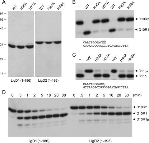
Figure 6.
End-healing activities of the AtuLigD1 and AtuLigD2 PE domains. (A) Aliquots (5 µg) of the Ni-agarose preparations of the wild-type (WT) and mutated versions of the N-terminal PE domains of AtuLigD1 and AtuLigD2 were analyzed by SDS-PAGE. The Coomassie blue-stained gel is shown. The positions and sizes (kDa) of marker polypeptides are indicated on the left. (B) Reaction mixtures (10 µl) containing 50 mM Tris-acetate (pH 6.0), 5 mM DTT, 0.5 mM MnCl2, 50 nM pmol 32P-labeled D10R2 primer-template (shown at bottom, with ribonucleotides highlighted in shaded boxes), and 4 µM WT or mutant PE domain as specified were incubated at 37°C for 20 min. The products were resolved by PAGE and visualized by autoradiography. The labeled species corresponding to the D10R2 substrate and the D10R1 end-product are indicated by arrowheads on the right. (C) Reaction mixtures (10 µl) containing 50 mM Tris-acetate (pH 6.0), 5 mM DTT, 0.5 mM MnCl2, 50 nM 32P-labeled D11p primer-template (shown at bottom) and 4 µM WT or mutant PE domain as specified were incubated at 37°C for 20 min. The products were analyzed by PAGE and visualized by autoradiography. The labeled species corresponding to the D11p substrate and the D11OH product are indicated by arrowheads on the right. (D) Reaction mixtures (90 µl) containing 50 mM Tris-acetate (pH 6.0), 5 mM DTT, 0.5 mM MnCl2, 100 nM 32P-labeled D10R2 primer-template and 4 µM AtuLigD1 or AtuLigD2 PE as specified were incubated at 37°C. Aliquots (10 µl) were withdrawn at the times specified above the lanes and quenched immediately with EDTA/formamide. The products were resolved by PAGE and visualized by autoradiography. The labeled species corresponding to the D10R2 substrate, the D10R1p intermediate and the D10R1 end-product are indicated by arrowheads on the right.

