
Figure 2.
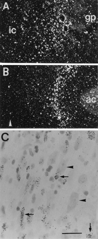
Survival and integration of transplanted neural stem cells. Dark-field images of autoradiograms of [3H]thymidine-labeled NGF-secreting cells grafted into the NBM or MS region (A and B). The cells had migrated out from the original implantation site and become integrated in the surrounding brain parenchyma (ic, internal capsule; gp, globus pallidus; ac, anterior commissure; arrowhead in B denotes the midline). (C) A bright field view of a Nissl-stained section at the MS target location, showing grafted cells (covered with silver grains, arrows) intermingled with host neurons (arrowheads). [Bar = 100 μm (A and B) or 15 μm (C).]