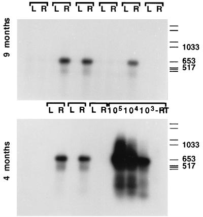
Figure 3.
RT-PCR amplification of the retroviral NGF transcript demonstrates long-term in vivo expression in samples from grafted animals. These animals received control- or NGF-secreting neural stem cells in the left (L) or right (R) hemispheres, respectively, and were sacrificed at 4 or 9 months postgrafting. Amplification of the retroviral transcript was performed on RNA extracted from frozen, dissected tissue blocks. The standards were prepared in parallel, by using known amounts of cells (103 to 105) taken from dividing cultures and mixed with rat brain tissue before RNA isolation. Lane labeled as −RT corresponds to the standard containing the highest amount of NGF cells, subjected to mock reverse transcription (in the absence of reverse transcriptase) and then run in parallel to the other samples for the PCR amplification. Molecular weight standards are Boehringer Type VI.

