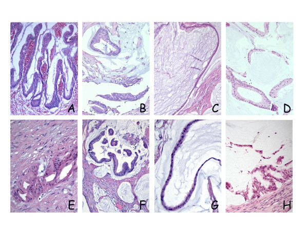
Figure 2.

Microscopic growth assessed by HE staining. Top panels (A, B, C and D) and bottom panels (E, F, G and H) represent sections from the PMP-1 and PMP-2 models, respectively. In both patients, primary tumor manifestations were appendiceal lesions; (A) Patient 1: cystadenoma of the appendix with low grade atypia. (E) Patient 2: mucinous adenocarcinoma, the selected section illustrating an area of invasive growth in the appendiceal wall. Peritoneal lesions from the main surgical specimens were remarkably similar in the two patients, exhibiting focal areas of cribriform growth and nuclear stratification (B and F), but in both cases, the histopathologic picture was dominated by strips of bland epithelium lining large accumulations of extracellular mucin (C and G). In xenografts from both models a similar histological growth pattern was observed, with adenomucinosis as the dominating manifestation, but with focal areas of nuclear stratification and cribriform growth, leading to classification as PMCA-I (D and H).
