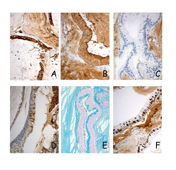
Figure 3.

Immunohistochemical and alcian blue staining of xenograft tissues. Consistently high membranous and cytoplasmic expression of CEA and CK20 (panels A and B, respectively) was observed in primary tumors, main surgical specimens and in all specimens harvested from the first six animal passages, here illustrated by sections from PMP-1 passage 1. CK7, on the other hand was hardly expressed in the PMP-1 series (panel C), whereas the PMP-2 model (passage 2) exhibited high expression of this cytokeratin (panel D), showing a distinct phenotypic difference between otherwise very similar tumors. Intra- and extracellular mucin was present in all examined sections (panel E). A high fraction of pKi67 positive cells was detected, and in this PMP-1 passage 3 tumor 10–50% of tumor cell nuclei were stained (panel F).
