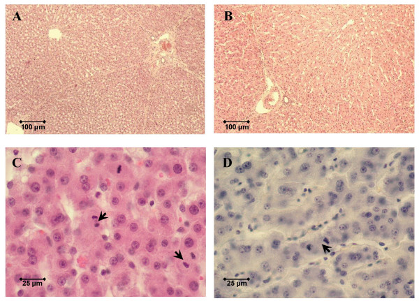
Figure 1.
Histology three days after surgery. Histology of pig livers three days after surgery was assessed to grade liver regeneration and hepatitis. Shown are representative low-power fields (original magnification 100×) in hematoxylin and eosin-stained sections of a control pig liver without surgery (A) and a pig liver three days after 70 to 80% hepatectomy (B). High-power fields (original magnification 400×) were used to count the number of mitotic hepatocytes (arrows) per ten visual fields. Representative microphotographs are shown for a pig liver three days after 70 to 80% hepatectomy (C) and a pig liver three days after 70 to 80% hepatectomy with hepatic decompression (D).

