Fig. 4.
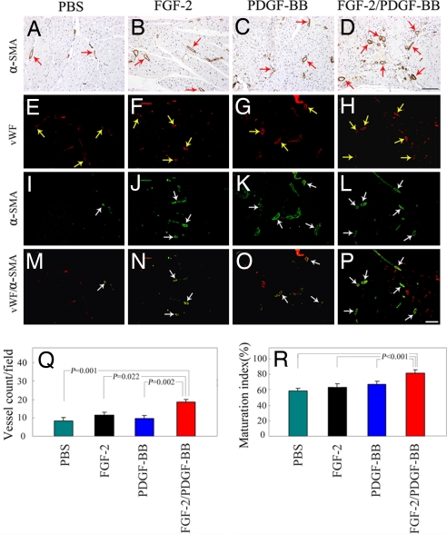
Immunohistochemical analysis of collaterals in the ischemic myocardium. An anti-α-SMA antibody was used to stain VSMC on paraffin-embedded (A–D) and frozen (green signals, I–L) sections of myocardium treated with single or combinatorial factors. The total number of blood vessels was revealed with an anti-vWF antibody (red signals, E–H), and the vWF/α-SMA double-positive vessel structures (yellow signals) were shown by superimposing both positive signals in the same sections (M–P). (Scale bars: 100 μm.) Myocardial collaterals were quantified from 10 random fields (×20 magnification), and the maturation index was quantified as the percentages of α-SMA-positive vessels vs. the total number of vessels (Q and R). The data are presented as means of determinants (±SEM).

