Fig. 5.
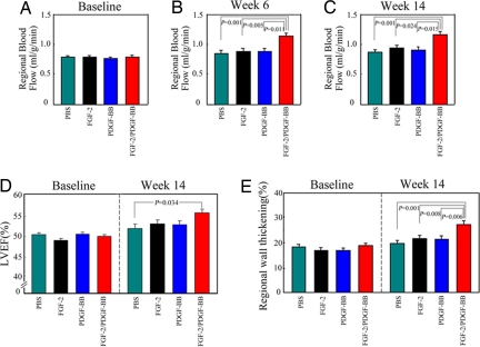
Regional blood flow and echocardiographic evaluation of global and regional cardiac functions. Regional blood flow was analyzed at day 0 (A), week 6 (B), and week 14 (C) by using a colored microsphere method, and the data are presented as mean values (±SEM) of the ischemic myocardial flow (ml/g/min). LVEF was measured to monitor the improvement of global function of the left ventricle after delivery of growth factors for 14 weeks (D). The values of regional wall thickening were obtained by using the open-chest echocardiography at day 0 and week 14 after treatments (E).

