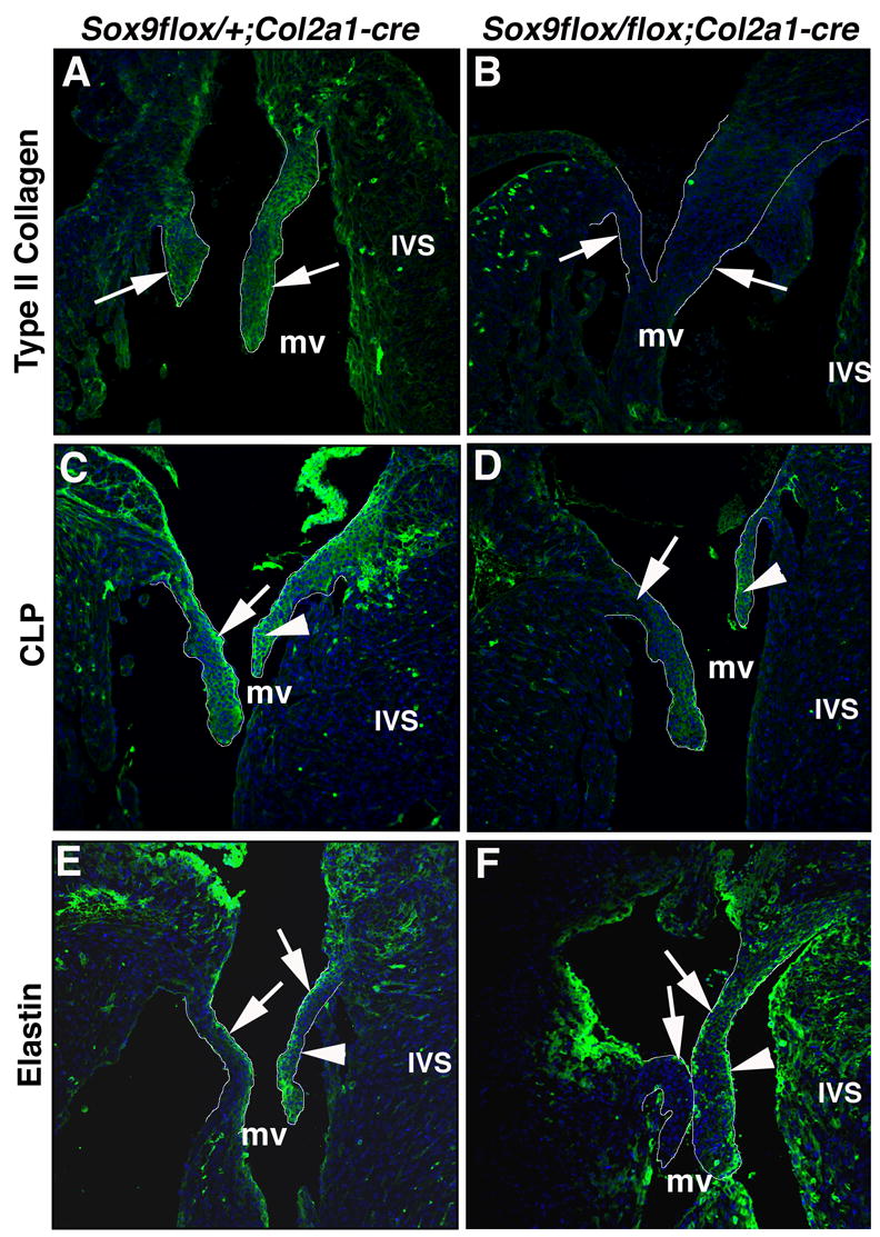
Figure 6. Type II collagen and CLP expression are reduced in Sox9flox/+;Col2a1-cre mice.

Immunostaining for type II collagen (A, B), CLP (C, D) and elastin (E, F) was used to determine ECM expression and organization in Sox9flox/+;Col2a1-cre (A, C, E) and Sox9flox/flox;Col2a1-cre (B, D, F) mitral valve mural and septal leaflets at E18.5 (outlined in white). The expression of type II collagen (A) and CLP (C) in the mural and septal valve leaflets (arrows) of Sox9flox/+;Col2a1-cre mice is diminished in Sox9flox/flox;Col2a1-cre mice (B, D). (E) Normal elastin expression is detected on the atrial surface of mitral valve mural and septal leaflets of Sox9flox/+;Col2a1-cre embryos. (F) Elastin expression is also observed on the atrial surface of valve leaflets from Sox9flox/flox;Col2a1-cre embryos, but ectopic expression is additionally noted on the ventricular surface of the septal leaflet (arrowhead, F). mv, mitral valve; IVS, interventricular septum.
