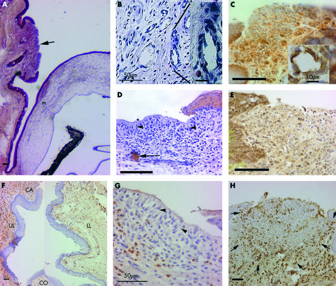
Figure 4.
(A) Location of leucocytic aggregate (arrow) in the palpebral conjunctiva (haematoxylin and eosin). Note goblet cells are absent in adjacent epithelium. m = graft margin. (B) Capillary displaying HEV morphology. (C) CD54 expression on basal epithelium, infiltrating cells, and capillary endothelium (inset). (D) Pancytokeratin staining showing lack of epithelium (note pallisading arrangement, arrowheads) overlying the aggregates but occasional cells (arrow) in the stroma. (E) MHC class I staining is not evident in superficial aggregate. (F) Composite picture showing CD4+ cells in upper (UL) and lower (LL) conjunctiva of the same section. Infiltration is more dense in UL, between the aggregates (CA) and the cornea (CO), than in LL. (G) NK cell are mainly in the stroma. Note pallisading (arrowheads) of cells in superficial layers. (H) ED2+ macrophages in circular arrangement in the aggregate defined by arrows. All unlabelled bars are 100 μm.

