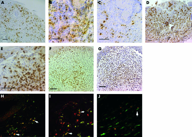
Figure 6.
(A) MHC class II expressing cells are numerous in superficial leukocytic aggregates. (B) CD4+ T cells colocalise with (C) CD25+ cells. (D) CD8α staining is greater in stromal areas and expressed on round and dendritic cells. (E) Granulocytes in aggregates within both superficial and stromal areas. (F) IFNγ expression and (G) TNFα expression in leucocytic aggregates. Immunofluorescence double staining showing cells (arrows) positive for (H) IFNγ (red) and CD8α (green) (×20); (I) IFNγ (red) and ED2+ macrophages (green) (×40); (J) TNFα (red) and ED2+ macrophages (green) (×40). Unlabelled bars are 50 μm.

