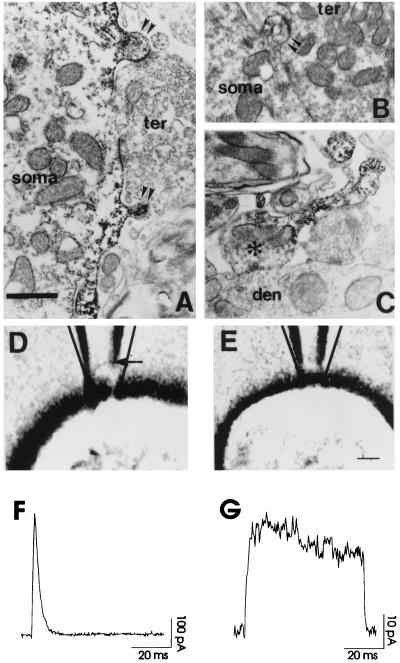
Figure 1.
Electron microscopic immunolocalization of Kv3.1 protein in restricted membrane compartments of auditory neurons. (A and B) Kv3.1 immunoreaction product found in the plasma membrane and underlying cytoplasm of spine-like protrusions (double arrowheads) of bushy cell somata (soma). The labeled spine protrusions are often present near or at the site of synaptic terminal (ter) contact. Note the difference in the neck geometry of the spine-like protrusions. (C) Photomicrograph of a Kv3.1 immunolabeled axon terminal (asterisk) (presumably from a bushy or MNTB neuron) synapsing on the dendrite of a parasuperior olivary neuron. Kv3.1 reaction product is concentrated in the neck region of the axon entering the synaptic terminal. [Bar = 0.75 μm (A and B), 0.91 μm (C)]. (D and E) A cell-attached vesicle or a vesicle-free patch following a seal in the patch pipette photographed through Nomarski optics. (Bar = 2.2 μm). (F and G) Currents recorded from the vesicle as in D and from the patch as in E. The calculated transmembrane potential was 140 mV (from −80 mV to +60 mV). The current density of CHO cells expressing Kv3.1 channels estimated from whole-cell recordings was about 10 pA/μm2, and single-channel conductance was 15 pS (14). This gave approximately 200 channels for the vesicle in F and 15 channels for the patch in G.

