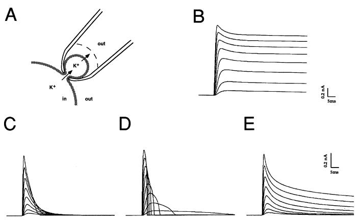
Figure 4.
Simulations of Kv3.1 current in a spine-like compartment. (A) Drawing of the geometry of the simulated configuration of the spine-like compartment, with potassium ion flux from the bulk cytoplasm into the head of the spine, and across the membrane into an unstirred layer on the surface of the external face of the membrane. (B) Simulations of Kv3.1 currents with fixed intracellular and intracellular potassium concentrations (120 and 3 mM, respectively). (C) Simulations of Kv3.1 current in a spine with a diameter of 0.9 μM, with restricted diffusion from bulk cytoplasm to spine and from the unstirred layer (thickness 50 nm) and bulk extracellular medium (kdiff and kext = 0, see Methods). (D) Simulations of Kv3.1 current in a spine of the same size with a fixed extracellular potassium concentration (3 mM, thickness of unstirred layer = 500 μm, kext =1.0) but with restricted diffusion from bulk cytoplasm to spine (kdiff = 0). (E) Simulations of Kv3.1 current with a fixed intracellular potassium concentration (120 mM) within the spine, but with restricted diffusion between the unstirred layer (thickness 50 nm) and bulk extracellular medium (kext = 0). Currents in all cases are shown in response to voltage steps from +10 mV to +150 mV in 20 mV increments.

