Figure 1.
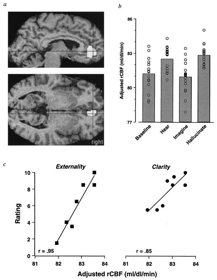
Anterior cingulate region activated in the group of hallucinators by both hearing and hallucinating conditions. (a) Projections on sagittal (upper) and transverse (lower) MRI templates of the region identified by the combination of four contrasts: the conjunction of two orthogonal contrasts (threshold, P = 0.001), hallucinate vs. baseline, and hearing vs. imagining, masked with two other orthogonal contrasts (threshold, P = 0.05), hearing vs. baseline, and hallucinate vs. imagining. The combination of these contrasts (with either pair masked by the other pair) isolates a significant region (P = 0.009, voxel level significance) that is activated by both hearing and hallucinating compared with both imagining and baseline conditions. This region of 207 voxels lies in the right ventral anterior cingulate, and its maximum (z = 4.60) is located at {6,48,0} (16); the crosshairs are located at these coordinates. (b) The adjusted rCBF response at {6,48,0} for each condition. The rCBF response is adjusted to an arbitrary mean of 50 ml/dl/min. Note the parallel rCBF changes during the hearing and hallucinating conditions. Subjects contributed two replications of each condition. (c) Correlations between the adjusted rCBF response at {6,48,0} and ratings of externality and clarity of the heard voices during the hallucinating condition. The average rCBF response and the average ratings of the two hallucinating conditions are plotted. The externality rating for one subject was unavailable.

