
Fig. 1.
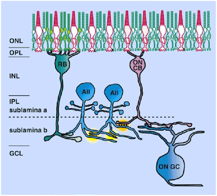
A schematic diagram of the role of the AII amacrine cell in the rod pathway. Each AII amacrine cell makes gap junctions not only with neighboring AII cells, but also with ON cone bipolar cells. These sign-preserving gap junctions with the bipolar cells distribute the signals from rod bipolar cells, which depolarize to light, into ON cone bipolar cells, which also depolarize to light. Sign-inverting inhibitory synapses from AII amacrine cells onto OFF cone bipolar cells occur in sublamina a (not shown), which serve to distribute the rod signal with the appropriate hyperpolarization to light into the OFF channel. The need for separate rod and cone ganglion cells is, thereby, avoided. ONL, outer nuclear layer; OPL, outer plexiform layer; INL, inner nuclear layer; IPL, inner plexiform layer; GCL, ganglion cell layer; RB, rod bipolar cell; CB, cone bipolar cell; GC, ganglion cell.