
Fig. 3.
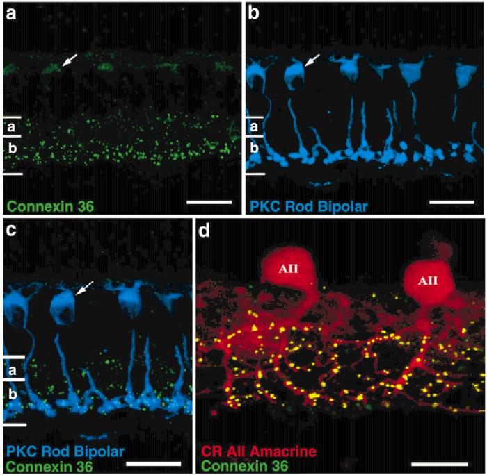
Cx36 immunoreactivity in oblique sections. a: Cx36 immunoreactivity appears as discrete puncta, densely in the ON region and more sparsely in the OFF region of the inner plexiform layer. Light labeling is also sometimes seen in the outer plexiform layer. A more diffuse cytoplasmic stain sometimes appears in rod bipolar cells (arrow). b,c: Staining of rod bipolar cells (blue) by an antibody to protein kinase C (PKC) demonstrates colocalization with the diffuse Cx36 immunostaining (green). d: Strong colocalization of Cx36 immunoreactivity (green) is seen on the processes in sublamina b of AII amacrine cells stained with anti-calretinin (CR, red). Light Cx36 immunoreactivity in sublamina a is not associated with the AII lobular appendages. Scale bars = 20 μm in a-c, 10 μm in d.